
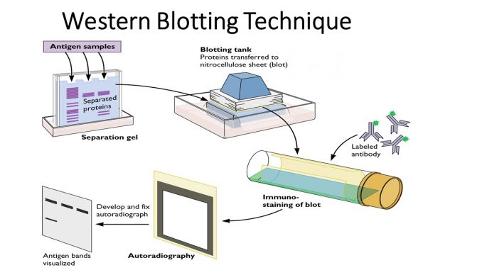
Once you’ve performed the western blot, including blocking, antibody incubations, and detection, the next crucial step is analyzing the protein bands on your membrane. Proper analysis ensures that your results are accurate, reproducible, and meaningful.
1. Visualizing Protein Bands
After the secondary antibody reacts with its substrate (commonly through chemiluminescence or colorimetric methods), protein bands appear on the membrane. The appearance and intensity of these bands correspond to the presence and relative abundance of the target protein.
-
Chemiluminescence: Enzyme on the secondary antibody reacts with a substrate, producing light captured by X-ray film or a digital imaging system.
-
Colorimetric detection: Produces visible colored precipitates at the site of the protein band, viewable directly on the membrane.
Tip: Capture images immediately or store them properly to avoid signal fading or background artifacts.
2. Comparing Bands to Molecular Weight Markers
Protein size is a critical parameter in western blotting. Most gels include a molecular weight ladder, which serves as a reference to estimate the size of your protein bands.
Steps:
-
Align the ladder bands with your target protein.
-
Compare the observed bands to the expected molecular weight of the protein.
-
Confirm specificity—unexpected bands may indicate cross-reactivity, degradation products, or post-translational modifications.
3. Quantifying Protein Expression
Western blots are often used for semi-quantitative or quantitative analysis of protein expression.
-
Densitometry Analysis:
-
Use imaging software (like ImageJ or commercial blot analysis tools) to measure band intensity.
-
The darker or more intense the band, the higher the protein concentration in that sample.
-
-
Normalization:
-
To account for unequal protein loading, normalize your target protein to a loading control, such as:
-
β-actin
-
GAPDH
-
Tubulin
-
-
Normalization ensures that differences in band intensity reflect actual differences in protein expression, not variations in sample loading.
-
-
Comparing Samples:
-
Quantified, normalized band intensities can be plotted for comparison between experimental conditions, treatments, or time points.
-
4. Detecting Non-Specific Bands
Sometimes, you may observe extra bands that do not correspond to your target protein. These may arise from:
-
Cross-reactivity of the primary antibody
-
Degraded protein fragments
-
Overloading the gel
Solutions:
-
Optimize antibody concentration
-
Use fresh samples and protease inhibitors
-
Adjust exposure times during detection
5. Considerations for Low-Abundance Proteins
For mechanism of western blotting expressed at very low levels:
-
Use high-sensitivity detection reagents.
-
Extend exposure time during imaging to capture faint bands.
-
Consider signal amplification techniques such as biotin-streptavidin systems.
6. Interpreting Band Patterns
Proper interpretation involves looking at both the size and intensity:
-
Single band at expected size: Confirms presence of the target protein.
-
Multiple bands: Could indicate isoforms, cleavage products, or non-specific binding.
-
Absence of band: Might mean the protein is not expressed under those conditions, or detection failed due to technical issues.
Always confirm your observations with positive and negative controls.
7. Tips for Reliable Analysis
-
Capture high-quality images to avoid saturation of strong bands.
-
Consistent exposure times allow accurate comparisons between blots.
-
Repeat experiments to confirm results, especially when quantifying changes.
-
Document everything: antibody concentrations, exposure times, and sample loading amounts for reproducibility.
Conclusion
Analyzing protein bands in a western blot is more than just looking at dark lines on a membrane—it’s about interpreting the presence, size, and relative quantity of proteins accurately. By carefully visualizing bands, using molecular weight markers, performing densitometry, and normalizing to loading controls, researchers can obtain reliable data on protein expression. Proper analysis not only validates experimental results but also ensures that conclusions drawn from western blotting are scientifically robust and reproducible.
